Rupture of the Cruciate Ligament
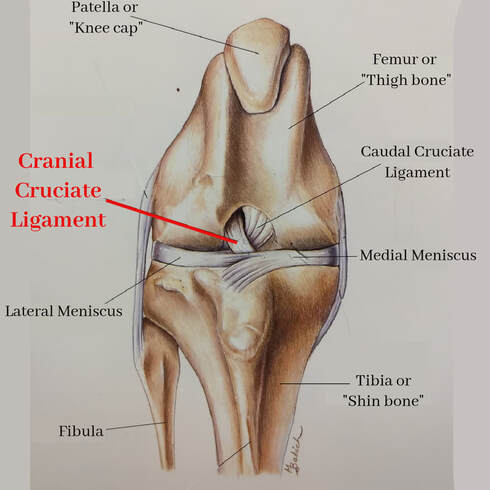
 Normal Stifle ("Knee") anatomy when viewed from the front.
The cruciate ligament is a tough fibrous rope of tissue within the knee joint (also called "the stifle") that gives the knee a hinge-like movement. This ligament is relatively easy to injure, especially if pets are active. Cruciate ligament injuries are common in people as well; sometimes described as "ACL tear" or a "football player's injury".
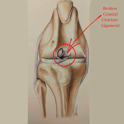
The knee joint in the dog (the second joint off the floor on the back leg) is relatively unstable because there is no interlocking of bones in this joint. Instead, the two main bones, the femur (thigh bone) and the tibia (shin bone), are joined with ligaments. There are two cruciate ligaments- the cranial cruciate (sometimes called the anterior cruciate) and the caudal cruciate (sometimes called the posterior). The cranial cruciate ligament is most often injured. The cruciate ligament can tear when the joint is twisted (twisting might occur when pets are quickly making a ninety degree turn or coming down from a jump at an awkward angle, for example). Once the ligament is torn, the bones of the joint can move past each other in an abnormal fashion and the pet will not want to bear much weight on the leg.
A torn cruciate ligament is diagnosed by demonstrating the abnormal motion of the tibia against the femur. This movement is called a "cranial drawer" sign. If your pet is very painful or has very strong leg muscles, this motion may be difficult to detect and your pet may benefit from pain relief or sedation. An xray of the leg may also be recommended. The cruciate ligament itself is not visible on xrays, but xrays will show inflammation inside the joint and allow evaluation of the bones of the joint.
|
Once the ligament is torn, the bones of the joint can move past each other in an abnormal fashion and the pet will not want to bear much weight on the leg.
A torn cruciate ligament is diagnosed by demonstrating the abnormal motion of the tibia against the femur. This movement is called a "cranial drawer" sign. If your pet is very painful or has very strong leg muscles, this motion may be difficult to detect and your pet may benefit from pain relief or sedation. An xray of the leg may also be recommended. The cruciate ligament itself is not visible on xrays, but xrays will show inflammation inside the joint and allow evaluation of the bones of the joint. |
Treatment for a torn cruciate ligament depends on many factors such as: pet size, pet activity level, concurrent joint disease, and if the pet is an anesthetic risk. Treatment options fall into two broad categories: medical management and surgical treatment.
Your Veterinarian will initially discuss medical pain management and exercise restriction (no off leash running or jumping, etc).
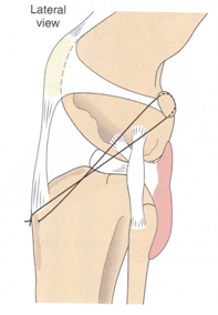
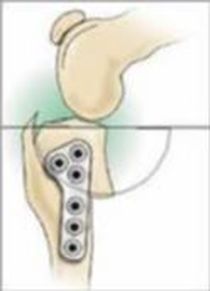
There are a few options for surgical therapy: all aimed at providing quick stability to the joint. Your surgeon will discuss which procedure they feel is best for your individual pet. One option is a "extracapsular procedure", during which a synthetic ligament replaces the body's broken one. Another procedure is called a TPLO or "Tibial Plateau Leveling Osteotomy". During a TPLO, the top portion on the tibia is cut away, rotated, and screwed back in place at a new angle that provides stability to the joint. With either surgical procedure, it is very important to follow post-operative directions very closely. Your surgeon will provide your pet with appropriate pain relievers as well as a physical therapy plan to get your pet back to normal movement after roughly 8 weeks.
Once surgery is performed, most pets recover relatively quickly. However, the development of arthritis is to be expected in any injured joint, so we strongly recommend glucosamine/chondroitin supplementation for all pets. Ask your veterinarian for a specific recommendation on joint products as they can vary greatly in effectiveness.
If surgery is not performed, the body will attempt to heal the joint instability by laying down scar tissue. This scar tissue takes many weeks to months to develop and your pet will likely have some loss of normal range of motion or persistent limp.


