Mast Cell Tumors
Mast cells are a type of immune system cell found normally in the skin, subcutaneous tissues, liver, lungs, and digestive tract. It is the job of mast cells to respond to allergens and parasites by secreting a variety of substances (heparin, histamine, and enzymes). When these cells multiply uncontrolled, they form a Mast Cell Tumor (MCT).
|
|
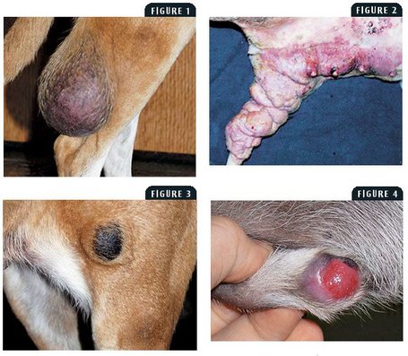
Mast Cell Tumors are one of the most common malignant skin cancers in dogs. MCT usually affects older pets although any age pet can technically develop these tumors. Any breed of dog can develop MCT, but certain breeds are predisposed: Boxers, Boston Terriers, Bulldogs, Pit Bulls, and Weimaraners are examples. MCT is somewhat uncommon in cats. MCT are sometimes called "the great imitators" because they do not have one characteristic appearance. MCT can form as skin masses of any size; they may be firm or soft, raised or flat, haired or ulcerated in appearance. These tumors can even fluctuate in size quickly- either shrinking or growing abruptly. This ability to change size is due to the substances secreted by the mast cells. Some MCT can actually appear as weeping sores that do not heal. |
Since mast cell tumors have a variety of appearances, accurate diagnosis requires obtaining a sample of the mass. A fine needle aspirate (FNA) is a diagnostic test in which a small needle is used to harvest cells from the mass which are then examined underneath a microscope. The size of the needle used for FNA is roughly the same size as that used for a vaccination, so this procedure is usually well tolerated by pets.
Depending on the type of mass being sampled, FNA is not always diagnostic, but, generally, mast cell tumors can be identified with this test.
Depending on the type of mass being sampled, FNA is not always diagnostic, but, generally, mast cell tumors can be identified with this test.
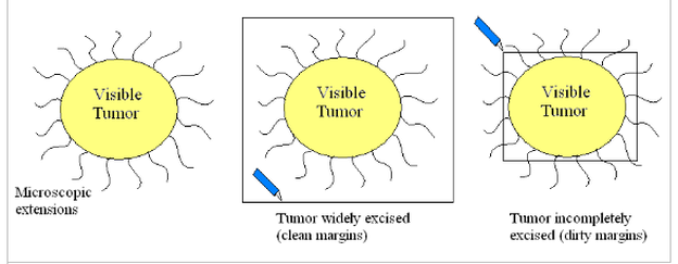
Treatment for MCT typically involve surgical removal. Aspiration and identification of the tumor type beforehand is important; however, as accurate diagnosis of a mast cell tumor will determine how much tissue needs to be removed during the procedure. MCT tend to make a visible mass or lesion on the skin surface, but also send out microscopic tendrils of cancer cells in all directions. Successful surgical excision of a MCT requires the removal of a large amount of normal appearing skin surrounding the tumor in an attempt to remove all cancer cells.
It is impossible to tell with the naked eye where these tendrils are and depending on the location of the tumor, complete surgical removal is not always possible.
It is impossible to tell with the naked eye where these tendrils are and depending on the location of the tumor, complete surgical removal is not always possible.
Your veterinarian may recommend bloodwork or other testing prior to surgery in order to look for spread of the tumor internally. When MCT spread or "metastasize", the cancer cells often travel to the liver, spleen, or lymph nodes. An anti-histamine, such as Benadryl, may be recommended before surgery also. An anti-histamine helps to combat the histamine that Mast Cells secrete- this may help reduce swelling and ulceration in preparation for removal of the mass. Some MCT shrink quite a bit with antihistamine treatment, but this does NOT mean that the cancer is gone!
After surgery, it is recommended that the removed tissue be sent to a laboratory for histopathological examination- this means that the whole mass will be examined microscopically to determine 1) if any cells where left behind in the pet and 2) what grade the MCT is. "Low Grade MCT" are present just in the skin and surgical excision is typically curative. Be aware, however, that new tumors may appear in the future on the same pet and they may be of different grades. "High Grade MCT" are more aggressive and metastasis to internal organs is more likely. Chemotherapy or other additional treatments may be recommended for these pets.
If complete surgical removal of the mass is not possible, your pet may be referred to a veterinary oncologist (cancer specialist) for further treatment including: advanced surgery with reconstruction, radiation therapy, and /or chemotherapy.


