Intervertebral Disk Disease (IVDD)
|
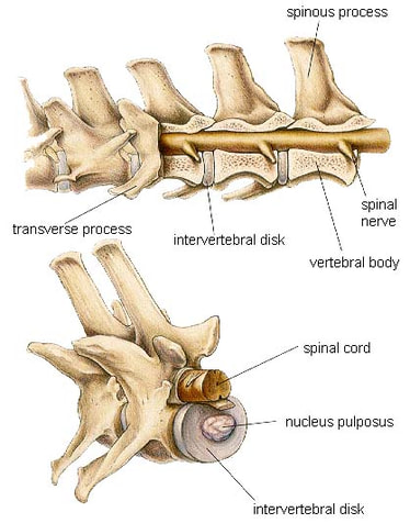
In order to explain intervertebral disk disease, we first need to review the normal anatomy of the spine (normal architecture of the spine is shown in the pictures to the left).
The spine is composed of individual bones called vertebrae that are separated by structures called intervertebral disks. These disks act like shock absorbers as your pet runs, jumps, and plays. Each disk is kind of like a jelly donut- it has a tough, round outer surface which is filled with a jelly-like goo called the nucleus pulposus. The spinal cord sits in a tunnel through the middle of all of the vertebrae. The spinal cord is a large bundle of nerves that relays instructions (nerve signals) from the brain down to the rest of the body. Your dog’s spine is divided into three regions: cervical, thoracic, and lumbar. In these three regions, the vertebrae have slightly different shapes, but the basic placement of the intervertebral disks and spinal cord is the same. The cervical spine supports your dog’s neck and shoulders. The thoracic spine supports the chest and abdominal region and the lumbar spine supports your dog’s lower back and hindquarters. Intervertebral disk disease (IVDD) occurs when the outer tough shell of the disk weakens, allowing the gelatinous center material escape. When this occurs, the intervertebral disk is said to have "ruptured" or "prolapsed". Since the outer shell of the disk is thinnest where it rests against the spinal cord, the leaking material is almost always expelled upward. At this point, the boney tunnel that normally protects the spinal cord becomes part of the problem as the spinal cord becomes compressed between the bone and the leaking disk material. |
|
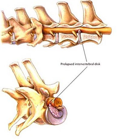
The picture to the right shows a ruptured intervertebral disk. Notice how the spinal cord is compressed by the escaping disk material. This compression and subsequent inflammation lead to intense pain and loss of spinal cord function.
Imagine the spinal cord is like a garden hose with nerve signals flowing from the brain down to the tip of the tail. If the "garden hose" is pinched in a certain location, the nerve signals may not get through "downstream", causing loss of nerve control to body structures beyond the pinch point. So far we have discussed intervertebral disk disease as if it just occurred in one location- disk ruptures can happen in multiple disks at once and symptoms will vary depending on the disk's location in the spine and whether the disk material compresses the spinal cord equally or off to one side. Symptoms of IVDD may include:
|
|
|
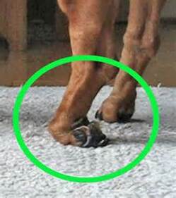
The image to the left shows a classic symptom of early IVDD- "knuckling over" of the foot. If you see your pet doing this- you NEED to see a veterinarian immediately! There is no way for you to treat this on your own at home and without intervention this symptom could progress to permanent paralysis.
"Knuckling over" is a sign that your pet has lost a innate reflex called conscious proprioception. Conscious proprioception is something that we all have. Close your eyes and put your hand out in front of you. Can you tell whether your hand is palm up or palm down without looking? Yes! You are using conscious proprioception! The nerves that handle conscious proprioception are located in the outer portion of the spinal cord, so if your pet knuckles over and stands on the foot as if they don't know that it isn't placed right- this is the first clue that spinal cord compression has started. |
Why does IVDD occur? The most common cause of IVDD is conformation and age. Over time, the intervertebral disks lose their flexibility, making them more susceptible to injury. Allowing your pet to become overweight puts excess strain on the spine and increases the risk of IVDD.
Occasionally, a known traumatic event will lead to IVDD. Many times, there is no inciting event- the pain seems to appear spontaneously, often in pets three to seven years of age.
Chondrodysplastic breeds (breeds with long backs and short legs) are most commonly affected due to the strain that dog's own anatomy puts on the spine. These breeds include: Dachshunds, Bassett hounds, Welsh Corgis, Beagles, Cocker spaniels, Pekingese, Poodles, and Bulldogs.
IVDD is diagnosed often by physical exam and xrays of the spine. Your veterinarian's exam will evaluate the pet for pain and try to localize where along the spine the ruptured disk is.
If you have one of the breeds listed above with acute back pain, IVDD is very likely. There are other diseases that can mimic IVDD such as: spinal embolism, bone fracture, degenerative myelopathy, and spinal cancer. If the diagnosis is in doubt, your pet may be referred to a specialist in order to perform advanced imaging such as a CT scan or fluoroscopy.
If you have one of the breeds listed above with acute back pain, IVDD is very likely. There are other diseases that can mimic IVDD such as: spinal embolism, bone fracture, degenerative myelopathy, and spinal cancer. If the diagnosis is in doubt, your pet may be referred to a specialist in order to perform advanced imaging such as a CT scan or fluoroscopy.
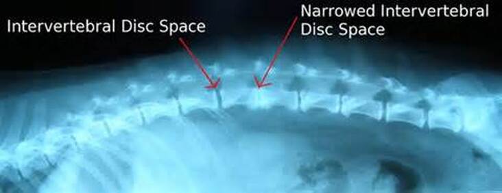
This is an xray of an IVDD patient.
The treatment for IVDD centers around pain control and preservation of spinal cord function. This may involve medical therapy or surgical intervention. To determine which therapies are needed, IVDD is designated into stages. Be aware that the stage of IVDD may change during treatment (even within hours to days)or can recur after treatment is completed.
Stage I- the pet is in mild pain and may recover after a few days without intervention. This stage may even go unnoticed by the pet owner.
Stage II- the pet is in moderate to severe pain and needs medication to be comfortable.
Stage III- the pet is partially paralyzed and is only walking with staggering or uncoordinated movements.
Stage IV- the pet has paralysis but still has the ability to feel the paralyzed limbs.
Stage V- paralysis and loss of feeling is present.
Pets in Stage II and III are usually treated medically with anti-inflammatories, pain relievers, muscle relaxers, and strict confinement of activity. Surgery may be considered if the pain or incoordination persists longer than 7 days or if the symptoms worsen from one day to the next. It is VITAL that confinement be enforced to avoid the pet exacerbating the disease.
Pets in Stage IV should have surgery, although a small percentage may recover without it. Dogs in Stage V should have surgery, and the sooner the surgery is completed, the better the prognosis (ideally, the surgery should be done within 24 hours of the onset of paralysis). Surgery is called a hemilaminectomy or a ventral slot surgery, depending on where the diseased disk is located in the spine. During the procedure, pieces of the vertebral bone are removed so that the leaking disk material can be extracted from around the spinal cord. Post-operatively, the pet often needs intensive care hospitalization to control pain, provide nutrition, and stimulate bladder function. Many weeks of physical rehabilitation follow surgery to help the pet regain function. In some cases, some neurologic dysfunction may be permanent.